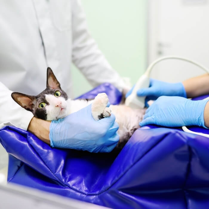
Abdominal Ultrasound

Abdominal Ultrasound
Ultrasound
Ultrasound is a pain-free, totally non-invasive technique that uses high-frequency sound waves to produce a real-time image of your pet’s internal organs. Often used in conjunction with radiography (x-rays), ultrasound provides a movie of what is happening inside your pet’s body. Additionally, unlike x-rays, diagnostic ultrasound does not use ionizing radiation, and there is no known health risk associated with its use.
Ultrasound is particularly useful in viewing your pet’s abdominal organs and evaluating heart functions. Abdominal ultrasound allows us to fully examine your pet’s liver, gallbladder, spleen, adrenal glands, pancreas, kidneys, urinary bladder, and parts of the stomach and intestines. Ultrasound also works well in conjunction with other diagnostic tools and a wide range of diagnostic procedures. For example, a radiograph of your pet’s abdomen may show enlargement of the liver but does not tell us why it is enlarged. An ultrasound allows us to see the liver’s structure in greater detail and identify specific lesions or masses. Using the ultrasound image as a guide, Alliston Veterinary Services veterinarians can obtain biopsies without major surgery, and your pet can often go home the same day.
Ultrasounds are typically not stressful for your pet and take anywhere from 30 to 60 minutes to perform. Dr. Christine Parker is certified in abdominal ultrasound and is able to perform an abdominal ultrasound, urogenital ultrasound, and pregnancy ultrasound in-house and submit images to a radiologist for interpretation. We also have a referral radiologist for cardiology consultations.

